BY DR TIM SANDLE | PHARMACEUTICAL MICROBIOLOGY AND CONTAMINATION CONTROL EXPERT
18th February
Vortex mixing is a common technique used in bioburden testing, especially in relation to the Total Microbial Aerobic Count (TMAC) method. This method is used as part of the pharmacopeial Microbial Limits Test (MLT) and used for intermediate sample testing during pharmaceutical manufacturing to assess bioburden levels as a process moves downstream.
The purpose is to ensure the thorough mixing of samples (homogeneity - to increase the representative nature of the tested subsample) and for detaching microorganisms from solid surfaces (the sampling container) into the sample. This process is necessary for ensuring that the subsequent microbial enumeration methods (like membrane filtration or plate counting) accurately reflect the total microbial load (bioburden) of the test sample.
This article focuses on the importance of vortex mixing.
Some microorganisms are more prone to clumping than others 1. Notable genera are the Staphylococci. Staphylococcus aureus is one of the most prominent bacteria known for clumping.
Clumping factors primarily refer to surface proteins (like ClfA and ClfB) from Staphylococcus aureus bacteria which is one of the most prominent bacteria known for clumping. It also secrets an enzyme called coagulase 2. While other Staphylococci (such as Staphylococcus epidermidis) grow in clusters, they lack the coagulase enzyme.
However, they can exhibit autoaggregation (clumping with their own kind) through other surface proteins and polysaccharides 3. Other bacteria prone to clumping include Escherichia coli 4 and some species of Bacillus, such as Bacillus subtilis and Bacillus cereus using similar mechanisms that enable them to form biofilms 5.
Bioburden sampling of incoming materials or during manufacturing is an important part of quality control. Additionally, bioburden determinations are important prior to sterilisation. While some bioburden sampling is of solid objects, the majority are likely to be from liquid samples. Bioburden refers to the population of viable microorganisms present on or in a product, component, raw material or environmental surface.
It is important to ensure that samples are representative and tested within a defined time, after being held under appropriate storage conditions. The sample itself is a subset taken from a whole population. Either the entire sample is tested or a subsample is drawn (a smaller portion taken from an existing sample, such as testing 1 mL using a pour plate method) 6.
As well as the prerequisites for processing, pre-test sample mixing is also an important.
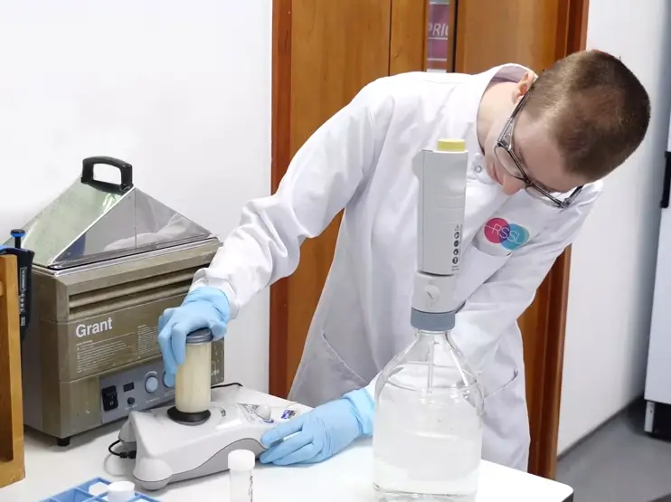
Samples should be mixed prior to testing. Given the inconsistencies of hand mixing, the most efficient way to achieve this is through vortex mixing 7. The primary purpose of vortex mixing in the bioburden testing procedure is to homogenise the sample and ensure the maximum possible recovery of viable microorganisms 8.
Vortex mixing relates to the vigorous blending of liquids in small containers (like test tubes, sampling universals etc) using a vortex mixer (or ‘vortexer’), a laboratory benchtop device that rapidly spins an off-centre rubber pad. As the motor runs, the rubber piece oscillates rapidly in a circular motion creating a whirlpool effect (the vortex) in the sample. Rotational speed settings range from 100 to 3,200 revolutions per minute (rpm). Vortex mixing is recommended for different microbiological methods 9, 10.
Uses of vortex mixing include:
Microbial extraction
When testing solid samples, such as medical devices or packaging materials, the item is submerged in a sterile extraction fluid (e.g., buffered water or 0.1% peptone water with a surfactant). Vortex mixing uses rapid, circular agitation to physically dislodge microorganisms from the product's surfaces and suspend them uniformly in the liquid.
Sample homogenisation
For both liquid and extracted samples, vortex mixing ensures that any microorganisms present are evenly distributed throughout the solution before an aliquot is taken for plating or filtration. This is essential for obtaining a representative count of the bioburden.
Dilution preparation
Vortex mixers are also used to resuspend stock solutions and ensure homogeneity when preparing serial dilutions of samples or microbial cultures, which is a standard procedure in quantitative microbiology.
The general procedure involves:
1. Sample preparation: The test sample is placed into a container with a measured amount of extraction fluid
2. Extraction: The sample is agitated using methods such as vortexing, sonication or mechanical shaking to remove microbes
3. Filtration/plating: An aliquot of the well-mixed extract is either filtered through a membrane (for filterable liquids) or mixed directly with culture media for plate counting
4. Incubation and enumeration: The plates or membranes are incubated under specified conditions (temperature and time) to allow microbial colonies to grow, after which the colonies are counted to determine the bioburden level
The effectiveness of the vortex mixing step is also necessary for the overall method suitability validation, which confirms that the chosen extraction method can reliably recover microorganisms from the specific product being tested. Hence, vortex mixing must be included in validation documents, with the time for vortex mixing and the rpm clearly stated.
Alternatives to vortex mixing include magnetic stirring and orbital shaking (for use with sample-solvent interaction for surface rinses). In some circumstances, these alternatives may yield improved recovery. In one study, time-course analysis showed significantly lower bacterial Colony-Forming Units (CFUs) with vortexing compared to sonication across all time points [11]. However, vortex stirring is generally a more useful method for routine work, combining convenience and rapidity of sample preparation with low running costs, low noise level and readily pipettable suspensions.
Whilst vortex mixing is important, samples for microbiological testing can be over mixed, which can potentially damage delicate cell structures and alter the results - the impact depends heavily on the specific application and the type of microorganism being tested. This is a reason why vortex mixing and the associated setting need to be included within the validation documentation.
The risk factors are:
Cell lysis or shearing
Vigorous or prolonged vortexing can physically shear the cell membranes or walls of fragile bacterial or fungal cells. This releases intracellular components, leading to an underestimation.
Altered viability
The physical stress from excessive agitation can kill or damage a portion of the microorganisms, leading to an underestimation of viable cell counts.
Protein denaturation
For assays involving sensitive enzymes or proteins, vigorous mixing can cause denaturation or foaming, reducing their activity and leading to inconsistent results.
Inconsistent results for pooled samples
The prolonged vortexing of pooled samples can lead to increased cell fragmentation and a decrease in cellular concentration.
As well as controlling speed and time, for applications where thorough mixing is needed but cell integrity is a concern, a pulse mixing mode (which cycles between mixing and pausing) can create a gentler alternative to continuous high-speed vortexing.
If the √N + 1 is flawed, why stick with it? The general answer is convenience. Traditional standards (ANSI/ASQ Z1.4, and ISO 2859) require large, resource-intensive sample sizes. In contrast, the √N + 1 approach delivers reduced test volume, ease of adoption and a reduction in the risk of accepting unsuitable material.
It all comes down to how much risk we are willing to accept and how much rigor we wish to put into the equation.
1. Fenchel, T. Microbial behavior in a heterogeneous world, Science 296(5570), 1068(2002)
2. Crosby HA, Kwiecinski J, Horswill AR. Staphylococcus aureus Aggregation and Coagulation Mechanisms, and Their Function in Host-Pathogen Interactions. Adv Appl Microbiol. 2016;96:1-41
3. Lowy FD. Staphylococcus aureus infections. The New England journal of medicine. 1998;339:520–532
4. Albert MJ, Qadri F, Haque A, Bhuiyan NA. Bacterial clump formation at the surface of liquid culture as a rapid test for identification of enteroaggregative Escherichia coli. J Clin Microbiol. 1993;31(5):1397-9
5. Di Franco C, Beccari E, Santini T et al. Colony shape as a genetic trait in the pattern-forming Bacillus mycoides. BMC Microbiol. 2002 13;2:33
6. Sanders, E. R. Aseptic Laboratory Techniques: Plating Methods. J. Vis. Exp. (63), e3064, doi:10.3791/3064 (2012).
7. A.N. Sharpe, Separation and concentration of samples, Detecting Pathogens in Food, 10.1533/9781855737044.1.52, (52-68), (2003).
8. Stocker, M Microorganisms in vortices: A microfluidic setup, Limnol. Oceanogr. 4, 392–398 (2006)
9. US FDA. Pharmaceutical Microbiology Manual, 2020: https://www.fda.gov/media/88801/download
10. ISO 11737-1:2018 Sterilization of health care products — Microbiological methods Part 1: Determination of a population of microorganisms on products, ISO, Geneva.
11. Arthur H. Totten, Li Xiao, Donna M. Crabb et al. Shaken or stirred?: Comparison of methods for dispersion of Mycoplasma pneumoniae aggregates for persistence in vivo, Journal of Microbiological Methods, 132, 2017: 56-62


